
Western Blot System
Western Blot System: Complete Scientific Guide for Protein Detection
What Is a Western Blot System?
A western blot system is a complete laboratory platform used to separate, transfer, detect, and analyze proteins from biological samples. It combines instruments, consumables, and detection tools required for the western blotting process, one of the most widely used techniques in molecular biology and biochemistry.
Scientists use a western blot system to identify specific proteins, compare expression levels, study signaling pathways, and validate experimental results.
Why Is a Western Blot System Important?
A reliable western blot system is essential because it allows researchers to:
Detect proteins with high specificity
Compare protein abundance between samples
Confirm gene expression changes
Study post-translational modifications
Investigate disease biomarkers
Validate proteomics findings
Generate reproducible laboratory data
This makes the western blot system a valuable tool in both research and clinical laboratories.
Main Components of a Western Blot System
1. Gel Electrophoresis Unit
This component separates proteins according to molecular weight using SDS-PAGE gels.
Key Functions:
-
Resolves protein mixtures
-
Produces clear band separation
-
Supports multiple sample lanes
2. Power Supply
The power supply delivers controlled voltage and current during electrophoresis and transfer.
Importance:
Stable electrical conditions are necessary for consistent protein migration and membrane transfer.
3. Protein Transfer Module
After separation, proteins are transferred from the gel to a membrane.
Common transfer methods include:
Wet transfer
Semi-dry transfer
Rapid dry transfer
Efficient transfer is critical for accurate protein detection.
4. Membranes
Proteins are immobilized onto membranes such as:
PVDF membrane
Nitrocellulose membrane
These membranes provide a solid surface for antibody binding.
5. Detection Reagents
Protein detection depends on:
Primary antibodies
Secondary antibodies
Chemiluminescent substrates
Fluorescent dyes
Colorimetric reagents
The quality of these reagents strongly influences final results.
6. Imaging Device
A western blot system often includes an imaging platform for visualizing protein bands.
Common imaging methods:
Chemiluminescence imaging
Fluorescence imaging
CCD camera systems
Digital gel documentation systems
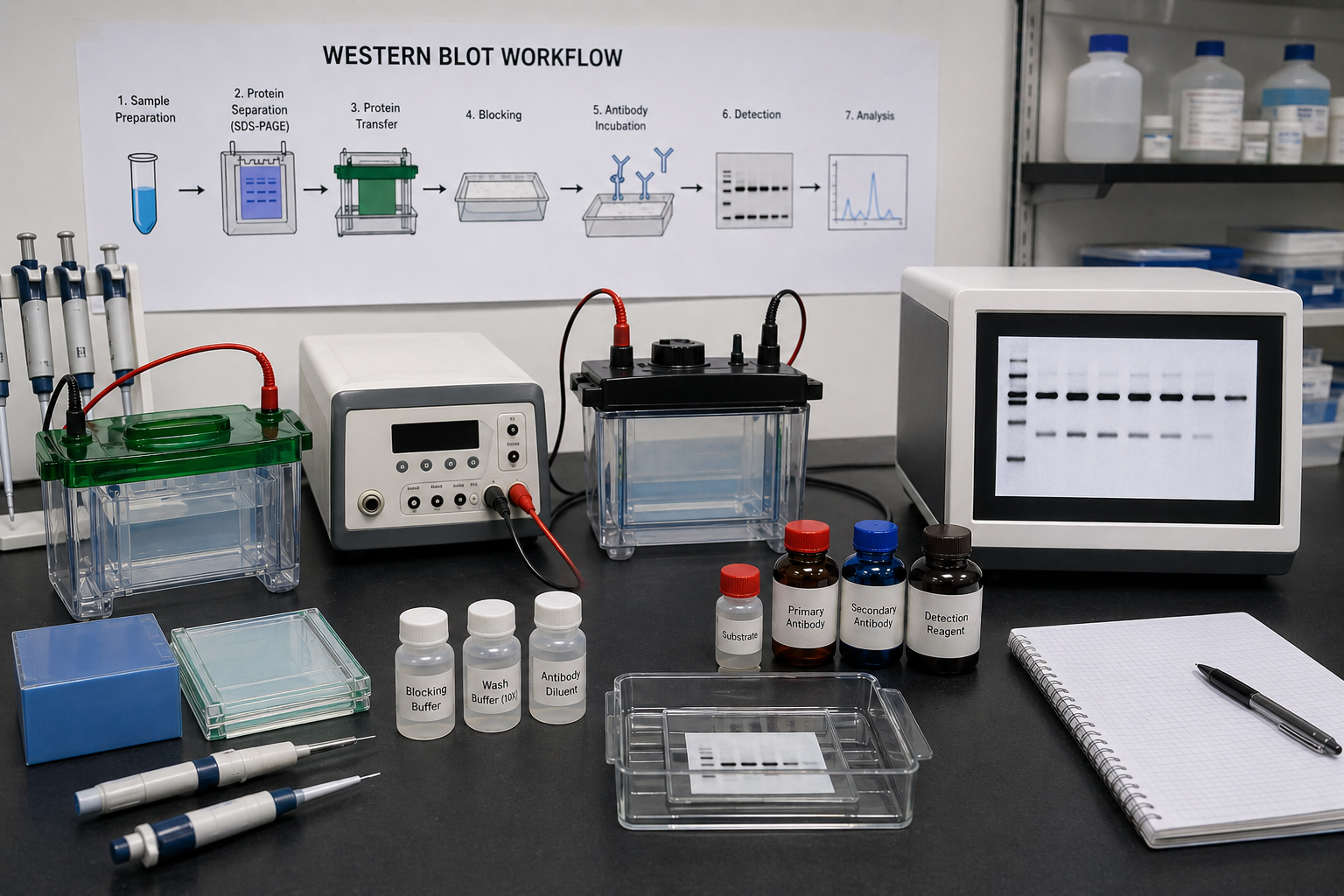
Western Blot System Workflow
Sample Preparation
Proteins are extracted from samples such as:
Cell cultures
Tissue homogenates
Blood fractions
Microbial cultures
Lysis buffers usually contain protease inhibitors to prevent protein degradation.
Protein Separation
Samples are loaded into polyacrylamide gels and separated by molecular weight.
Protein Transfer
Separated proteins are moved onto a membrane using electrical current.
Blocking
The membrane is incubated with blocking solution to reduce non-specific antibody binding.
Antibody Incubation
The membrane is treated with:
-
Primary antibody specific to the target protein
-
Secondary antibody linked to a detection label
Signal Detection
Bands corresponding to the target protein are visualized and recorded.
Data Analysis
Band intensity may be quantified to compare relative protein expression levels.
Types of Western Blot System
Manual Western Blot System
Uses separate instruments for each step.
Advantages:
-
Flexible protocol design
-
Lower initial equipment cost
Semi-Automated Western Blot System
Combines faster transfer and imaging technologies.
Advantages:
Reduced hands-on time
Improved consistency
Automated Western Blot System
Uses integrated or capillary-based platforms.
Advantages:
Minimal manual handling
High reproducibility
Quantitative performance
Applications of Western Blot System
Cancer Research
Detection of tumor markers and signaling proteins.
Neuroscience
Analysis of synaptic and neuronal proteins.
Immunology
Measurement of immune-related proteins and cytokines.
Stem Cell Biology
Monitoring differentiation markers.
Drug Discovery
Validation of target engagement and pathway modulation.
Infectious Disease Research
Detection of microbial or host-response proteins.
Common Problems and Troubleshooting
Weak Bands
Possible causes:
Low protein loading
Inefficient transfer
Weak antibody binding
High Background
Possible causes:
Excess antibody concentration
Poor washing steps
Inadequate blocking
Uneven Bands
Possible causes:
Air bubbles during transfer
Uneven gel polymerization
Inconsistent voltage
How to Improve Western Blot Results
Use fresh samples when possible
Keep proteins cold during extraction
Measure protein concentration accurately
Optimize antibody dilution
Confirm transfer efficiency
Include positive and negative controls
Use proper washing procedures
Future of Western Blot System Technology
Modern western blot system technology continues to evolve with:
Faster transfer devices
High-sensitivity imaging systems
Multiplex fluorescent detection
Automated workflows
These innovations improve speed, sensitivity, and reproducibility.
Final Thoughts
A western blot system remains one of the most trusted tools for protein detection and analysis. From sample preparation to image acquisition, every component plays a role in producing accurate and reproducible data.
Whether used in academic research, biotechnology, or diagnostics, the western blot system continues to be a cornerstone technique for studying proteins and biological pathways.
Recent Posts
-
How to Inactivate Proteinase K
How to Inactivate Proteinase K: Complete Guide for Molecular Biology Labs Introduction In molecular …5th May 2026 -
Western Blot System
Western Blot System: Complete Scientific Guide for Protein Detection What Is a Western Blot System? …27th Apr 2026 -
Biocoupler™ Bundle
Biocoupler™ Bundle – Save Costs and Enhance Culture Performance Maximize the efficiency of the paten …23rd Apr 2026